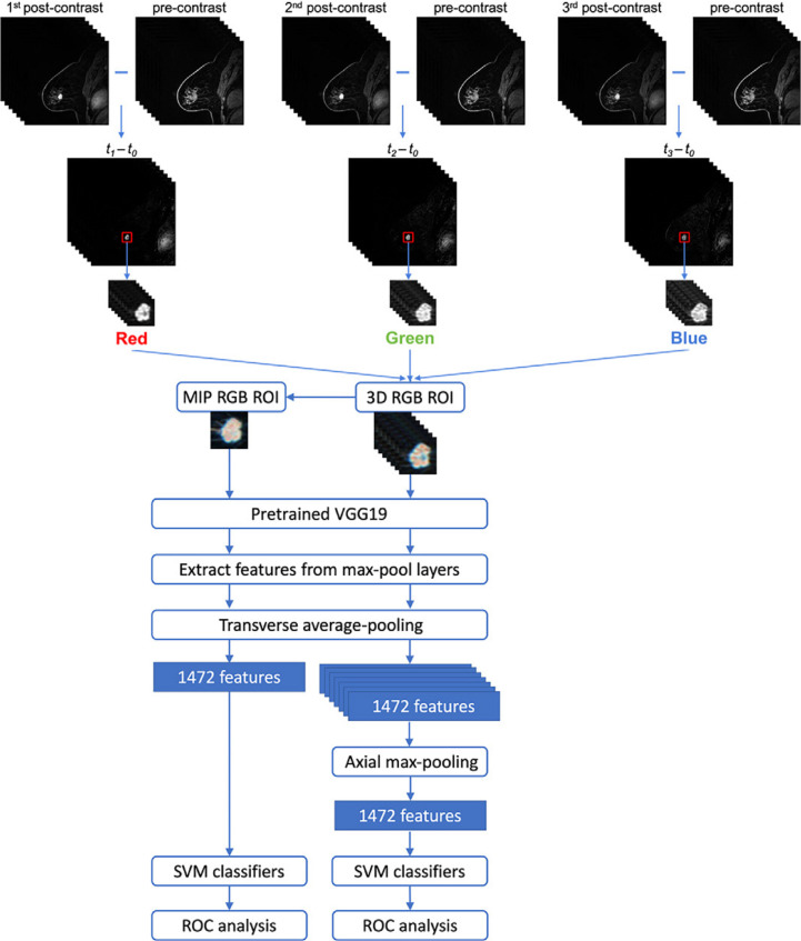
Figure 5.
An illustration of combined DL feature extraction methods with traditional machine learning classifiers for lesion classification from cropped ROI’s of MIP and 3D RGB of subtraction images of DCE MRI sequences. 74 The top layer illustrates the construction of the cropped ROI’s from the DCE MRI sequences. MIP and 3D RGB features were integrated with max-pooling and then passed to a machine learning classifier. Reprinted by permission from Radiology: Artificial Intelligence, 74 copyright Radiological Society of North America 2021. DCE, dynamic contrast-enhanced; DL, deep learning; MIP, maximum intensity projection; ROI, region of interest.

