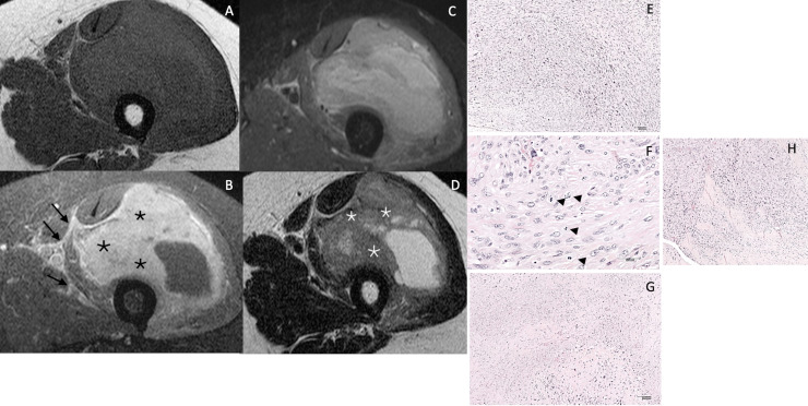
Figure 3.
65 year old male patient with important swelling of the left thigh. T1 (A), contrast enhanced FS T1 (B), STIR (C), T2 weighted sequence (D). Major intratumoral enhancement (≥ 51% of tumour volume (black asterisks), peritumoural enhancement (arrows) and heterogenous signal on STIR, indicating high grade tumour. Internal areas of low T2 signal (white asterisks). Surgical biopsy revealed a high-grade leiomyosarcoma grade III by FNCLCC Intersecting fascicles of pleomorphc spindle cells (E-Hematoxylin and eosin, H&E, original magnification × 100), with high rate of mitotic figures (arrows) (F- H&E, original magnification × 400), and large areas of necrosis (G- H&E, original magnification × 100). Infiltrating aponevrotic tissu in the periphery of the tumor (H- H&E, original magnification × 100)

