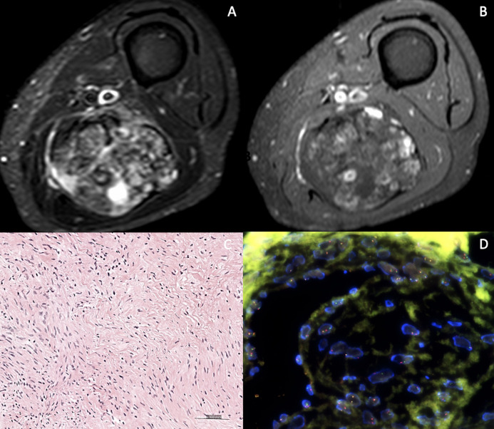
Figure 4.
86 year old female patient with a palpable mass of the posterior lower left thigh discovered on MRI. STIR (A) and contrast enhanced FS T1 (B). Highly heterogenous mass on STIR, but tumoural enhancement <51% of the total volume (arrows) and no peritumoral enhancement. Surgical biopsy revealed a low grade fibromyxoid sarcoma (C- Hematoxylin and eosin, H&E, original magnification × 200) Scattered hyperchromatic cells admixed with heavily collagenized areas. No mitoses nor necrosis were seen. (D- By FISH- Fluorence in situ hybridization) FUS Dual Color Break Apart Probe detected a translocation involving the FUS gene.

