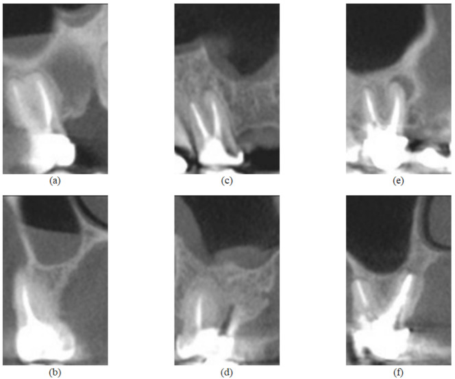
Figure 2.

Feature images from the UFPE database, where (a,b) represent a tooth without lesion and are sagittal and coronal sections, respectively; (c,d) represent a tooth with small lesion and are sagittal and coronal sections, respectively; (e,f) represent a tooth with large lesion and are sagittal and coronal sections, respectively.
