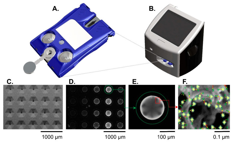
Figure 2.
The Smart Immunoassay platform hardware consists of a cartridge (A) and a portable instrument (B). The instrument activates blister packs on the cartridge, performs the multistep immunoassay, and collects the immunofluorescent signal from the agarose beads. Panels (C–F) show the sensor(s) at different length scales. A scanning electron micrograph (C) shows the microfluidic cartridge’s sensor matrix without beads. A fluorescent image shows the same sensor matrix with beads present (D). Out of the 20 beads in the sensor matrix, a single agarose bead (encircled in green dotted line) is magnified (E) and shows a strong immunofluorescent reaction signal against a dark background. Panel (F) is a further magnified view of an agarose bead and illustration representing the fluorescent immunocomplexes formed on agarose bead fibers. The immunocomplexes are in sandwich configuration with capture antibodies (green symbols), antigen (yellow symbols), detecting antibodies (red symbols), and fluorophore (glowing yellow symbols). Reproduced from [20] with permission from the Royal Society of Chemistry.

