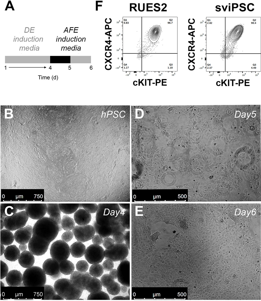
Figure 1. Differentiation of hPSC to AFE.
A. Schematic representation of the differentiation protocol until day 6. B-E. Bright-field views after culture of RUES2 ESC to confluence (B), to DE (C) and to AFE (D,E) (representative of n>10 independent experiments). F. Representative example of the flow cytometric profile after staining day 4 RUES2 ESC and sviPSC DE cells for CXCR4 and cKIT (representative of n>10 independent experiments). DE, definitive endoderm; AFE, anterior foregut endoderm.

