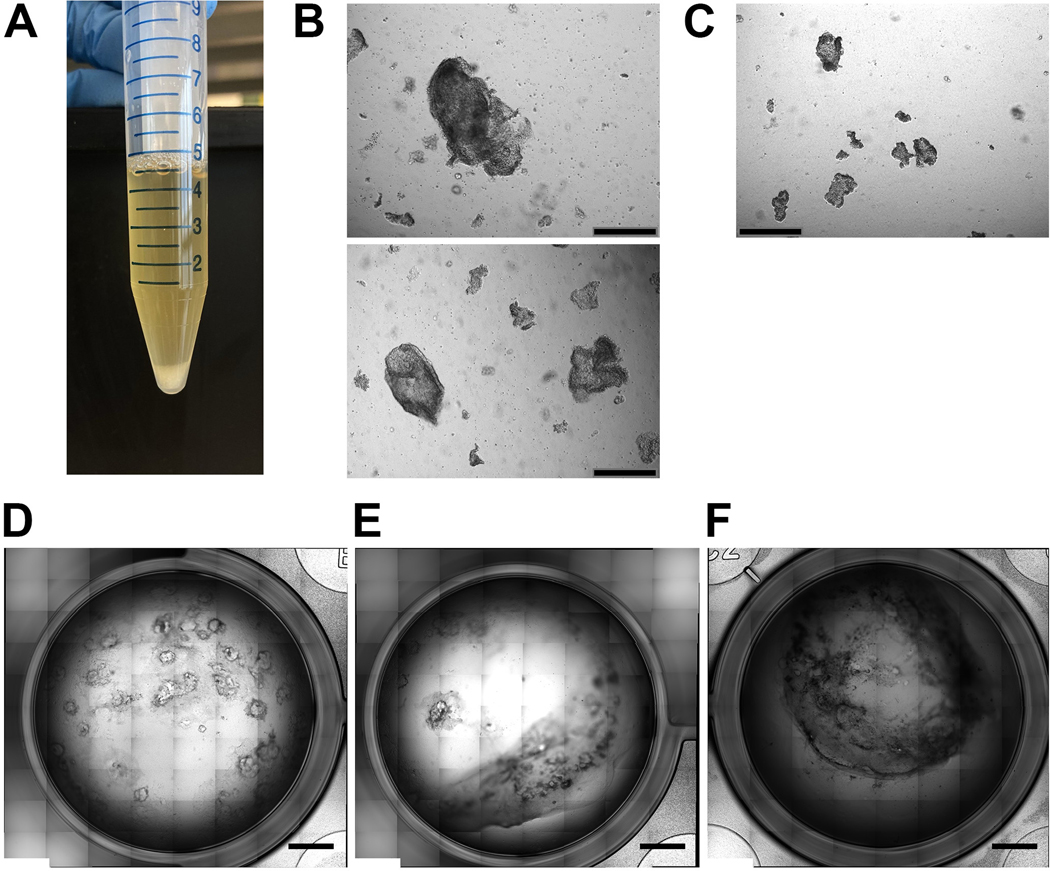
Figure 3. Practical aspects of the culture protocol.
A. Representative example of what to expect when disrupting the cell monolayer into cell clumps for day 15 and day 25 replating (steps 35 and 50). The clumps will pellet to the bottom of the conical tube and the media will be cloudy from single cells/very small cell aggregates in suspension. B. Representative example of the bright-field appearance of appropriately sized cell clumps immediately after replating into collagen I gel (steps 49–52) (scale bar 500μM). C. Representative example of the bright-field appearance of small sized cell clumps that exhibit lower survival rate upon replating immediately after replating into collagen I gel (steps 49–52) (scale bar 500μM). D-F. Potential appearances of the collagen I layer over the course of the differentiation. (D) Intact collagen layer at day 50 of the differentiation protocol; (E) Collagen layer partially detached and retracted from the well wall; (F) Contracted collagen I gel (D-F scale bar 2.5mm).

