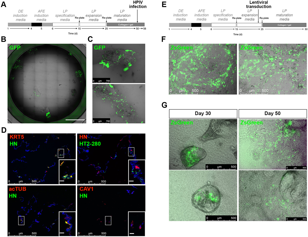
Figure 9. Applications of the differentiation protocol.
A. Schematic representation of HPIV3 infection of day 50 cells differentiated according to our protocol. B. Bright-field view and GFP expression in a representative culture well of day 58 RUES2 ESC differentiated according to the protocol shown in A and infected with GFP expressing-HPIV3 at day 50 (n=3 independent experiments, scale bar: 5mm). C. Higher magnification bright-field views and GFP expression in the cultures 8-days post infection with GFP expressing-HPIV3 (representative of n=3 independent experiments). D. Immunofluorescence staining for viral particle HN and mature airway and distal lung markers 8 days post-infection with HPIV3 (representative of n=3 independent experiments, inset scale bar: 30μm). E. Schematic representation of lentiviral transduction of lung progenitors before embedding in collagen I gels and further differentiation and maturation according to our protocol. F. Representative bright-field views and ZsGreen expression in lung progenitors 44h after transduction with a ZsGreen expressing-lentivirus, according to established protocols (representative of n=3 independent experiments). G. Representative bright-field views and ZsGreen expression of transduced lung progenitors further cultured in collagen I gels according to the protocol shown in A, at the indicated differentiation days (representative of n=3 independent experiments). DE, definitive endoderm; AFE, anterior foregut endoderm; LP, lung progenitor.

