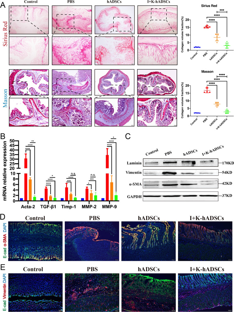
Fig. 3.
The IFN-γ- and KYNA-primed hADSCs significantly inhibited ECM deposition and EMT process. (A) Collagen staining analysis for therapeutic efficacy evaluation of colon fibrosis in rats; Up: Sirius Red staining; Bottom: Masson’s trichrome staining and Staining area statistics by the ImageJ software (n = 5); (B) qPCR analysis of fibrosis-related gene expression (Acta-2, TGF-β1, Timp-1, MMP-2, and MMP-9) in the colon on 46th day after treatment (n = 5); (C) Western blot analysis of laminin, vimentin, and α-SMA protein expression; (D) Immunofluorescence staining of the epithelial marker E-cad and myofibroblasts marker α-SMA in frozen sections of the colon (Scale bar, 100 μm); (E) Immunofluorescence staining of the epithelial marker E-cad and stromal marker vimentin in frozen sections of the colon (Scale bar, 100 μm); Data are represented as mean ± SEM

