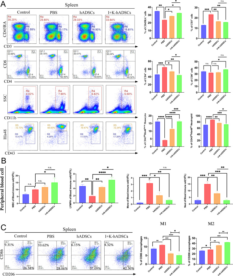
Fig. 4.
The IFN-γ- and KYNA-primed hADSCs can promoted the polarization of M2 macrophages. (A) Rat with the TNBS-induced colon fibrosis were killed on day 46 to harvest the spleen, mLN (n = 3). CD45RA+ B cells, CD3+ T cells, CD4+ T cells, CD8+ T cells, CD43+His48Int−Lo monocytes and CD43+His48Hi neutrophils in spleens of each group were detected by flow cytometry and statistical (Int-Lo means interval lower positive cells, Hi means strongly positive cells); (B) Blood routine analysis of WBC, lymphocyte, monocytes, and neutrophil in peripheral blood after primed hADSCs treatment (n = 3); (C) Analysis of macrophage subsets change. The infiltration of CD86+M1 macrophages and CD206+M2 macrophages in spleens of each group was detected; the following statistical figure shows the analysis of CD86+M1 and CD206+M2 macrophages in the CD68+ cell population (n = 3); data are represented as mean ± SEM

