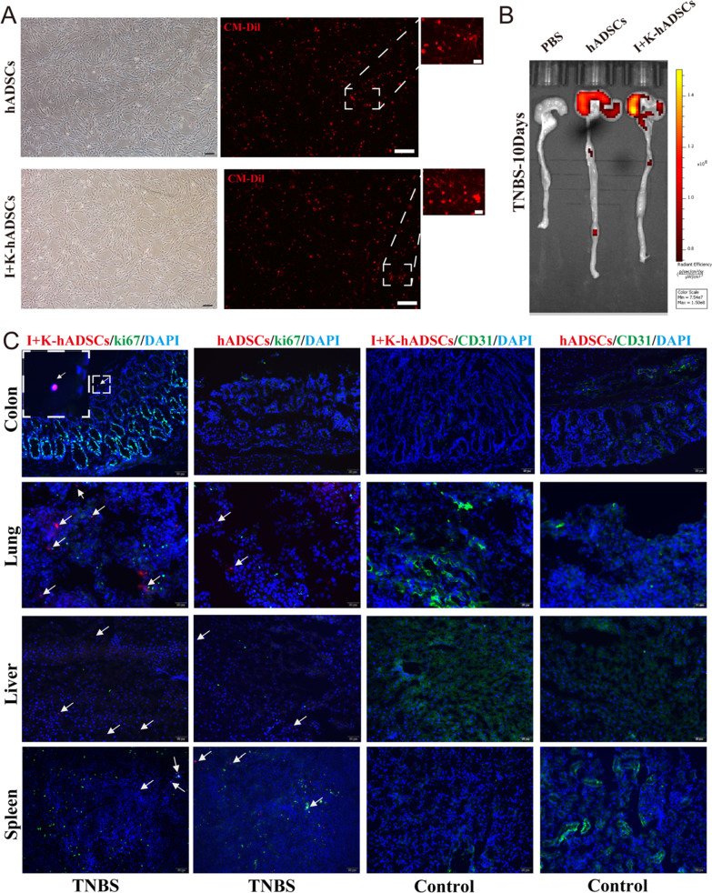
Fig. 6.
The IFN-γ and KYNA improved hADSCs homing to the colon tissue. (A) The morphology of Dil-labeled hADSCs and primed hADSCs was observed under microscope (scale bar, respectively, 50, 100, 10 μm); (B) The rats were killed on day 10 and the fluorescence signals of Dil-labeled cells in the colon and small intestine were observed under the IVIS system; (C) In vivo distribution of hADSCs and primed hADSCs was observed by immunofluorescence staining of the colon, lung, liver, and spleen. The white arrow points to the Dil-labeled hADSCs (Scale bar, 50 μm)

