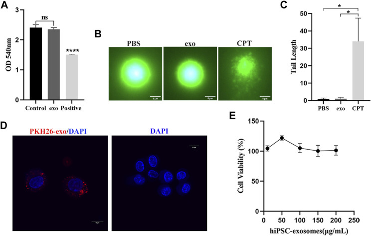
FIGURE 3.
Safety evaluation of hiPSC-exosomes at the cellular level. (A) Absorbance values of various groups of erythrocyte suspensions at 540 nm. N = 4 per group (ns, no statistical difference; ****p < 0.0001). (B) Fluorescence image of DNA after alkaline comet electrophoresis of leukocytes in PBS, hiPSC-exosomes, and the 50 μM camptothecin group. (C) Tail length analysis of comets in the three groups. N = 5 per group (*p < 0.05). (D) PKH26-labeled hiPSC-exosome uptake by RAW264.7 cells under laser scanning confocal microscopy. (E) Viability of RAW264.7 cells treated with up to 200 μg/ml hiPSC-exosomes. N = 4 per group. Data are expressed as the mean ± SEM.

