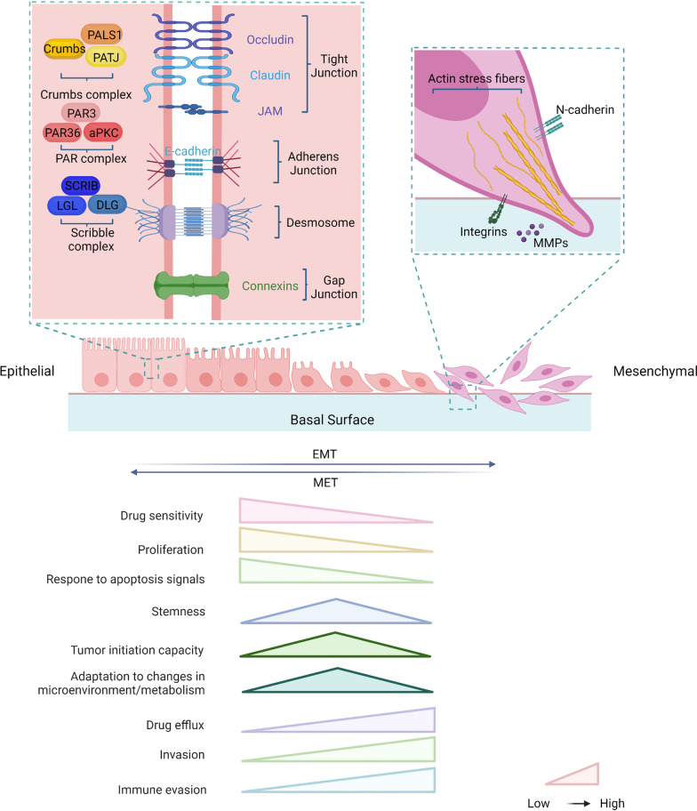
Fig. 1.
Changes in the cytoskeleton and properties between epithelial and mesenchymal during the EMT process. Epithelial cells exhibit apical–basal polarity with cell–cell and cell–matrix attachment. Three multi-protein complexes (Scribble complex, Crumbs complex, and PAR complex) interact to regulate the spatial separation of apical and basal structural domains together to establish cell polarity. Intercellular adhesion and communication are provided by intercellular junctions and maintain tissue stability and integrity. Tight junctions (TJs) form strips around cells that help separate apical and basal regions and form sealed spaces between adjacent cells, preventing the flow of material. Adherens junctions (AJs) are located below TJs, surround cells, and provide intercellular adhesion, but they are relatively permeable. Gap junctions are gaps located on the outer surface of cells and are hydrophilic ion transport channels between adjacent cells. Bridging granules provide sites of cell adhesion and intermediate filament binding to disklike structures located on the outer surface of the cell. The occurrence of EMT leads to the dissolution of intercellular junctions and loss of cell polarity allowing cytoskeletal rearrangements that alter the shape of the cell, transforming the cell into a mesenchymal phenotype and promoting cell motility and invasion. Based on a synthesis of the literature, we conclude that as the EMT progresses, the cell characteristics are changed, including reduces in drug sensitivity, proliferation, and response to apoptosis signals and increases in drug efflux, invasion, and immune evasion. The partial EMT with intermediate state has properties of enhanced stemness and tumor initiation capacity, and stronger ability to adapt to the changes in immune microenvironment and metabolism

