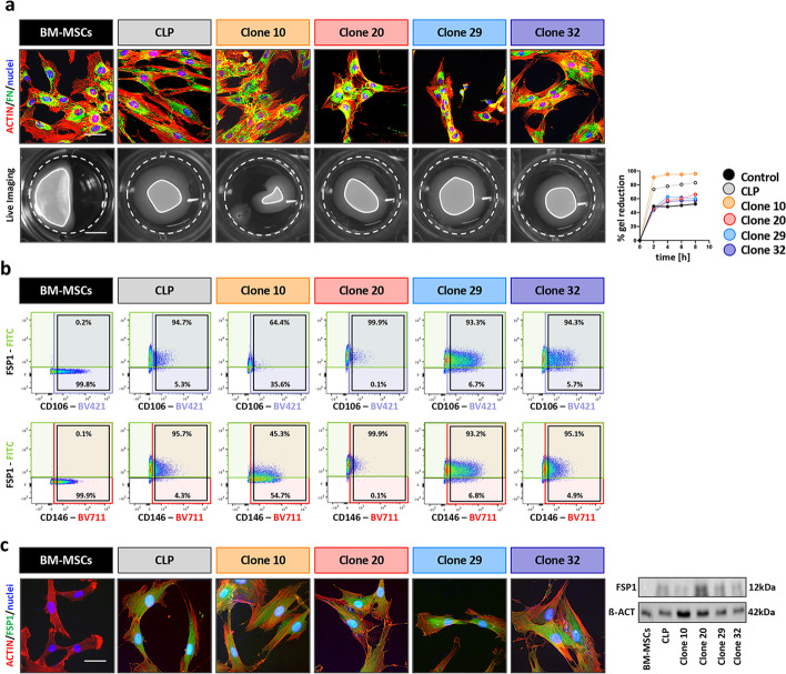
Fig. 6.
a IF images of BM-MSCs, CLP and Clones 10, 20, 29 and 32 for human FN (green). Scale bar: 20 µm. Actin (red); nuclei (blue). Live imaging pictures of detached collagen gels seeded with BM-MSCs, CLP and Clones 10, 20, 29 and 32 (time:8 h). White dashed lines indicate the initial area of the gels, while the white lines indicate the final size of the gels. Scale bar: 7.5 mm. Progressive gels reduction was measured by ImageJ over a period of 8 h and plotted in the diagram on the right. b Scatter plots display the co-expression of CD106 and FSP1 (top row), or of CD146 and FSP1 (bottom row) in BM-MSCs, CLP and Clones 10, 20, 29 and 32. CD106, CD146 and FSP1 positive cells are included in the purple, red and green boxes, respectively. Black boxes indicate CD106 or CD146 populations. Percentages indicate the fraction of CD106 or CD146 positive cells that are positive (top) or negative (bottom) for FSP1 expression. Gating strategy is presented in Additional file 1: Fig. S6. c IF staining and immunoblot for FSP1 protein expression confirmation in BM-MSCs, CLP and Clones 10, 20, 29 and 32. Full-length blots are presented in Additional file 1: Fig. S5. Scale bar: 20 µm. FSP1 (green); actin (red); nuclei (blue). kDa: kilo Dalton

