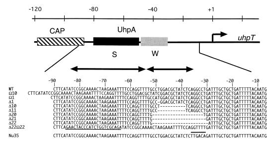
FIG. 2.
Promoter mutations. The schematic representation of the uhpT promoter shows the location and extent of protein-binding regions along the top. CAP, the CAP-binding site; S, the higher-affinity UhpA-binding region; W, the weak or lower-affinity UhpA-binding region. The sequence changes in the promoter variants used in this study are depicted along the bottom. Deleted residues are indicated by dashes. The 22-base insertion in Δ22 Ω22 is underlined, and the sequence changes to introduce a consensus −35 sequence in the Nu35 variant are indicated by an overbar.

