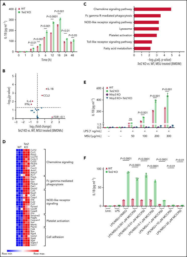
Figure 4.
Loss of Tet2 augments MSU crystal-induced secretion of IL-1β in macrophages. (A) Time-course analysis of IL-1β levels in supernatant of WT and Tet2 KO BMDMs treated with an MSU crystal dose of 100 µg/mL. P values were obtained using 2-sample Student t test. (B) Cytokine array in supernatant of Tet2 KO vs WT BMDMs after 6 hours of treatment with MSU crystals. Red dots highlight cytokines with FDR < 0.1. (C) KEGG pathway enrichment analysis of differentially expressed genes in Tet2 KO vs WT BMDMs treated with MSU crystals. (D) Heatmap of selected differentially expressed genes in inflammatory pathways from Tet2 KO vs WT BMDMs after administration of MSU crystals. (E) Dose-response analysis of IL-1β levels in supernatant of MSU crystal-treated BMDMs obtained from WT, Tet2 KO, Nlrp3 KO, and Tet2 KO+Nlrp3 KO mice. P values were obtained using 2-sample Student t test. (F) Dose-response analysis of IL-1β levels in supernatant of MSU crystal-treated BMDMs incubated with MCC950. BMDMs were obtained from WT and Tet2 KO mice. P values were obtained using 2-sample Student t test.

