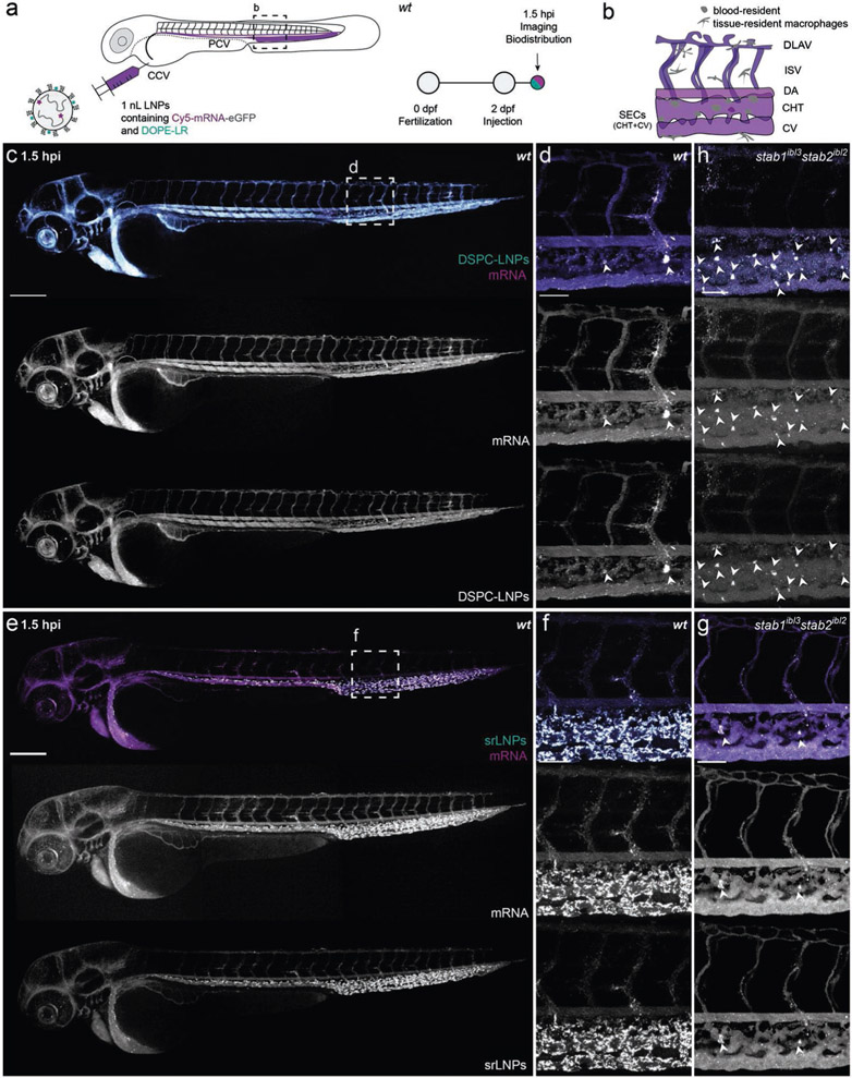
Figure 2.
Biodistribution of DSPC–LNPs and srLNPs in two-day old embryonic zebrafish at 1.5 hpi. a) Schematic showing the site of LNP injection (i.v.) within embryonic zebrafish (2 dpf) and imaging timeframe. LNPs contained DOPE-LR (cyan, 0.2 mol%) as fluorescent lipid probe and Cy5-labeled eGFP mRNA (magenta) as fluorescent mRNA probe. Injected dose: ≈10 × 10−3 m lipid, ≈0.2 mg kg−1 mRNA. Injection volume: 1 nL. Major venous blood vessels: CCV: common cardinal vein; PCV: posterior cardinal vein. b) Tissue level schematic of a dorsal region of the embryo containing scavenging cell types (i.e., SECs and blood resident macrophages). Blood vessels: DA: dorsal aorta, CHT: caudal hematopoietic tissue; CV: caudal vein; ISV: intersegmental vessel; DLAV: dorsal longitudinal anastomotic vessel. c,d) Whole embryo (10× magnification) and tissue level (40× magnification) views of DSPC–LNP biodistribution within wild-type (AB/TL) embryonic zebrafish (2 dpf) at 1.5 hpi. DSPC–LNPs were mostly freely circulating, confined to, and distributed throughout, the vasculature of the embryo. Low level phagocytotic uptake within blood resident macrophages is highlighted with white arrowheads. e,f) Whole embryo (10× magnification) and tissue level (40× magnification) views of srLNP biodistribution within wild-type (AB/TL) embryonic zebrafish (2 dpf) at 1.5 hpi. srLNPs were mainly associated with SECs within the PCV, CHT, and CV of the embryo and were largely removed from circulation at 1.5 hpi. Phagocytotic uptake of both DSPC–LNPs and srLNPs within blood resident macrophages at 1.5 hpi was confirmed by analogous LNP injections in transgenic mpeg:mCherry zebrafish embryos, stably expressing mCherry within all macrophages (see Figure S1 in the Supporting Information). g) Tissue level (40× magnification) view of srLNP biodistribution within stab1−/−/stab2−/− mutant zebrafish embryos[56] at 1.5 hpi. Within stabilin KOs, srLNPs were now mostly freely circulating, with low level phagocytotic uptake within blood resident macrophages highlighted by white arrowheads. h) Tissue level (40× magnification) view of DSPC–LNP biodistribution within stab1−/−/stab2−/− mutant zebrafish embryos[56] at 1.5 hpi. Within stabilin KOs, DSPC–LNPs remain mostly freely circulating, with low level phagocytotic uptake within blood resident macrophages highlighted by white arrowheads. For whole embryo images of LNP biodistribution within stab1−/−/stab2−/− mutant zebrafish embryos[56] at 1.5 hpi, please see Figure S2 in the Supporting Information. Scale bars: 200 μm (whole embryo) and 50 μm (tissue level).

