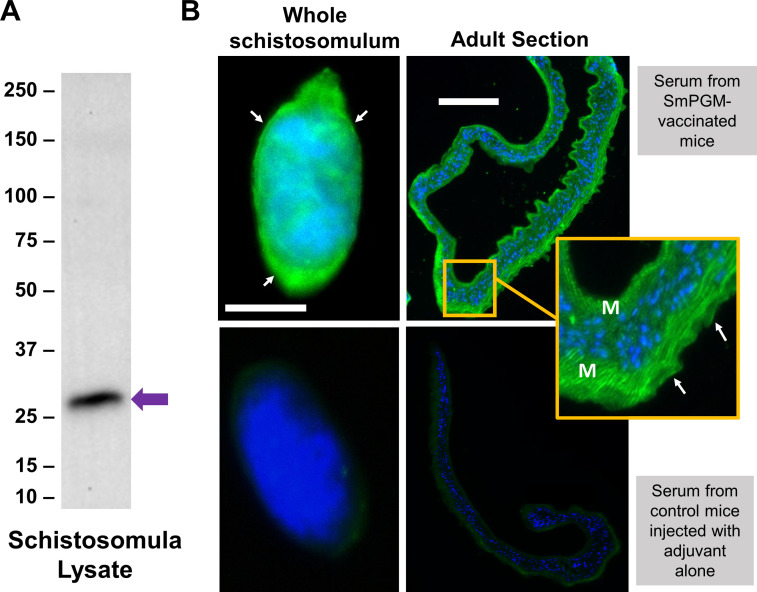
Figure 1.
Immunolocalization of SmPGM in Schistosoma mansoni schistosomula and adult worms. (A) Western blot analysis demonstrating binding of antibodies from rSmPGM-immunized mice to a protein of the expected size of native SmPGM in a Schistosoma mansoni schistosomula lysate (arrow). Blots probed with sera derived from mice immunized with adjuvant alone showed no binding to parasite lysate (not shown). Numbers to the left indicate markers representing molecular mass in kDa. (B) Indirect immunofluorescent labeling of native SmPGM (green) in a whole fixed S. mansoni schistosomulum (top left) and a section of an adult S. mansoni worm (top right) using sera from mice immunized with rSmPGM. Tegument staining is indicated by white arrows. Sub-tegumental muscle staining is indicated (M) in the inset yellow-bordered box. Counterstaining with DAPI highlights nuclei (blue). Lower images show a schistosomulum (left) and an adult section (right) incubated with serum from mice injected with adjuvant alone as negative controls. Scale bars: 100 μm.

