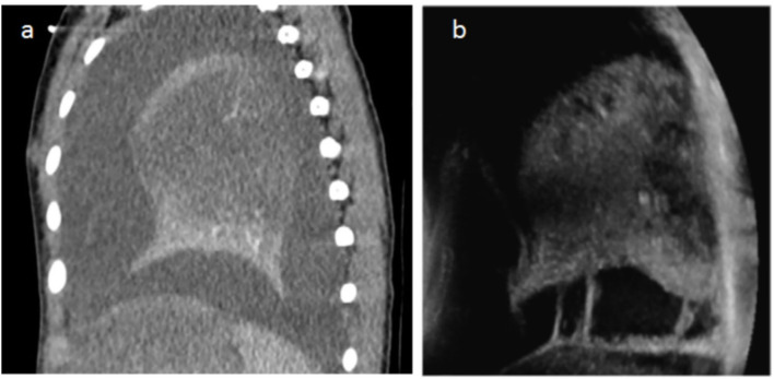
Figure 3.
(a) Chest CT with contrast agent injection in a 2.5-year-old child with 39°C fever, cough, and breathing difficulties. Sagittal reconstructions show a heterogeneous lung with unenhanced parenchyma corresponding to necrosis and a large pleural effusion. The US performed the same day also shows heterogeneity and hypodensity of the parenchyma in the necrotic zones (same as on CT) and pleural effusion. The periphery of the lower lobe is spared in a similar way on LUS and CT. In addition, US demonstrates bands of fibrin within the effusion, not visible on CT (b).

