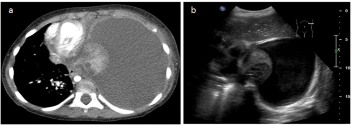
Figure 4.
A 3-year-old female patient with severe dyspnea. (a) The transverse slice chest CT demonstrates massive left effusion and atelectasis of the entire left pulmonary parenchyma, with heterogeneous enhancement and round necrotic unenhanced lesions posteriorly. (b) LUS performed the same day demonstrates the same parenchymatous damage with coalescent cystic lesions corresponding to necrosis.

