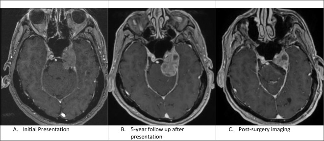
Figure 1.
A: Axial C+ T1-weighted image shows an enhancing mass centred in the left cavernous sinus with mild displacement of the medial left temporal lobe. B: Axial C+ T1 weighted image shows interval enlargement of the prepontine component of the mass. There are also some new cystic changes within the mass. C. Axial C+ T1-weighted image shows residual enhancing mass lesion after resection of the cisternal component of the mass.

