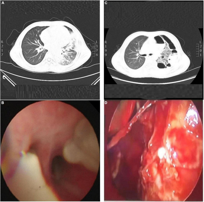
FIGURE 1.
(A) CT scan of his chest, show the left lung and the right lower lobe consolidation, with atelectasis in the lower lobe of the left lung. The left pleural cavity showed a small amount of pleural effusion, and the lumen of the bronchial branch of the left lower lobe was not unobstructed. (B) Bronchoscopy, show the basal segment of left lower lobe, the bronchial mucosa was rough, a large number of yellow and white mucus plugs were found in the opening, and the ventilation was not smooth. (C) Chest CT scan. Indicate consolidation of left and right upper and lower lobes with signs of atelectasis and multiple cavities in the left lung, partially wrapped left pneumothorax. (D) Thoracoscopy, show the lung surface covered with a yellow purulent moss-like layer.

