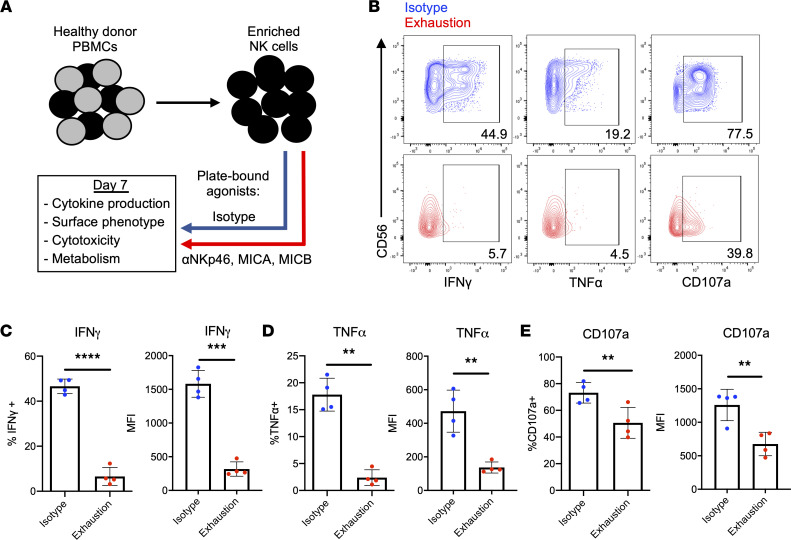
Figure 1. Prolonged stimulation through activating receptors induces NK cell exhaustion.
(A) Schematic representing the in vitro model of exhaustion. Agonists of NKp46 (anti-NKp46) and NKG2D (MICA and MICB) were adsorbed onto tissue culture plates and used to stimulate NK cells for 7 days. Plate-bound isotype IgG served as a control. Both groups received 1 ng/mL IL-15. (B) NK cells harvested from isotype-coated and exhaustion plates (day 7) were incubated with K-562 targets for 4 hours (E/T: 2:1). Cytokine production (IFN-γ and TNF-α) and degranulation (CD107a) were measured via flow cytometry. Isotype NK cells (top row) in blue, exhausted NK cells (bottom row) in red. E/T, effector/target. (C–E) Quantification of cytokine production and degranulation as percentage of parent population (live, CD3–CD56+ cells) and mean fluorescence intensity (MFI) (n = 4). Paired t tests were used for comparisons. **P < 0.01; ***P < 0.001; ****P < 0.0001.

