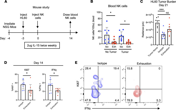
Figure 5. Prolonged stimulation through activating receptors results in functional and proliferative defects in vivo.
(A) Schematic representing experimental design: sublethally irradiated NSG mice were injected (i.v.) with HL-60-Luc2 cells and injected (i.v.) with NK cells 3 days later. NK cells had been incubated for 7 days on plates with either isotype IgG (control) or anti-NKp46 and MICA/B as previously described. (B) Fourteen days after NK cell injection, blood was drawn, and NK cells were counted via flow cytometry. NK cells were human CD45+CD56+CD3–. One-way ANOVA was used for comparisons (n = 8). *P ≤ 0.05. (C) HL-60 tumor burden was tracked via bioluminescent imaging (BLI), with day 21 data pictured. One-way ANOVA was used for comparisons (n = 8). ***P < 0.001. (D and E) Fourteen days after NK cell injection, NK cells were restimulated with K-562 leukemia cells for 4 hours as previously described. Ki67, IFN-γ, TNF-α, and CD107a expression was assessed via flow cytometry (D). Paired t tests were used for comparisons (n = 8). *P ≤ 0.05. Representative data depict Ki67 and IFN-γ expression following K-562 restimulation (E).

