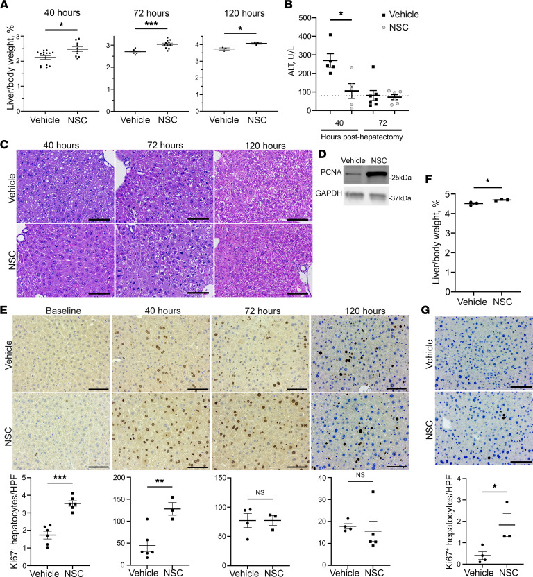
Figure 1. NSC-87877 accelerates liver regeneration following murine partial hepatectomy.
(A) Mean liver/body weight ratio in mice treated with vehicle or NSC (7.5 mg/kg/dose, twice daily) 40 hours (n = 10-15), 72 hours (n = 6-11), and 120 hours (n = 3) after hepatectomy. (B) Plasma alanine aminotransferase (ALT) (U/L) 40 and 72 hours after hepatectomy (n = 5-7) in mice treated with vehicle or NSC. Dotted line represents the upper limit of normal (79 U/L). (C) Representative H&E-stained liver sections 40, 72, and 120 hours after hepatectomy in vehicle- and NSC-treated mice. Scale bar: 100 μm. (D) Proliferating cellular nuclear antigen (PCNA) immunoblot from murine whole liver lysates, 40 hours after hepatectomy, with GAPDH as a loading control. Representative of image of 2 independent experiments. (E) Representative Ki67 IHC-stained liver sections in vehicle- and NSC-treated mice from baseline, 40, 72, and 120 hours after hepatectomy (n = 3) quantified in 10 HPF (200×) per section. Scale bar: 100 μm. (F) Mean liver/body weight ratio in vehicle or NSC-treated mice following 4 weeks of treatment (n = 3). (G) Representative Ki67 IHC-stained liver sections in vehicle- and NSC-treated mice after 4 weeks of treatment. Ki67+ hepatocytes were quantified in 10 HPF (200×) (n = 3). Data are shown as mean ± SEM (*P < 0.05, **P < 0.01, ***P < 0.001). Statistical analysis was performed with 2-tailed Student t test.

