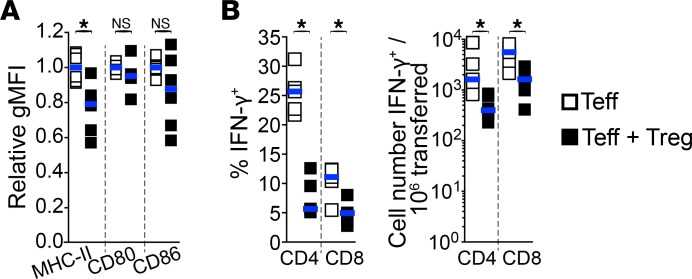
Figure 5. Tregs reduce expression of both MHC-II on APCs and IFN-γ in Teffs within allografts.
Innate immune cells were characterized by flow cytometry from islet allografts of mice lacking SLOs (as described in Figure 1A), 7 days after cell transfer. (A) Tregs significantly reduce MHC-II, but not CD80 and CD86, expression levels on CD11c+MHC-II+ innate cells within allografts. Relative mean geometric MFI of MHC-II, CD80, and CD86 expression levels on live CD45+Lin–CD11c+MHC-II+ cells within islet allografts. n = 4–6 mice per group from 2–3 independent experiments. (B) Tregs reduce IFN-γ expression in Teffs within allografts. Percentage (left) and absolute cell numbers (right) of IFN-γ–expressing CD4+ and CD8+ Teffs by direct in vivo cytokine assessment. n = 5 per group from 2 independent experiments. Each square represents data from 1 mouse and horizontal bars show median. Mann-Whitney tests used. *P < 0.05.

