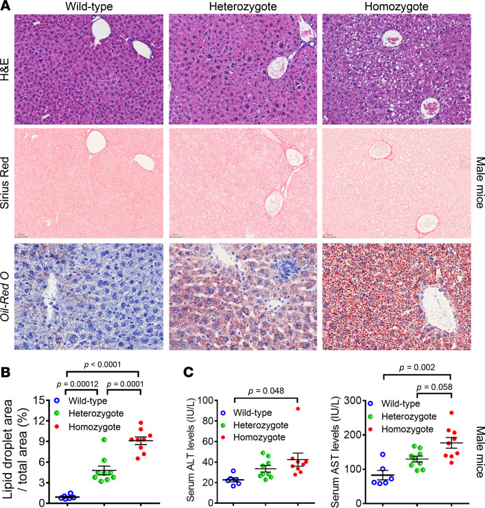
Figure 2. The Sema7aR145W mutation causes intrahepatic accumulation of small lipid droplets in male mice at 10 weeks of age.
(A) Representative images (original magnification, ×200) of H&E staining, Sirius red staining, and Oil Red O staining in WT and Sema7aR145W heterozygous and homozygous male mice at the age of 10 weeks. (B) Analysis of lipid droplets in the Oil Red O–stained liver sections of WT (n = 6) and Sema7aR145W heterozygous (n = 9) and homozygous male mice (n = 9). (C) The levels of serum ALT and AST in Sema7aR145W WT (n = 6), heterozygous (n = 9), and homozygous male mice (n = 9). The data were analyzed by 1-way ANOVA with Tukey’s post hoc tests or by Kruskal-Wallis test with Dunn’s post hoc test analysis.

