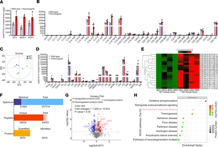
Figure 3. The Sema7aR145W mutation increases hepatic FA and TG concentrations in mouse livers.
Male WT and Sema7aR145W homozygous (HO) mice at 10 weeks old (n = 4 per group) were euthanized and their liver samples were prepared. (A) Gas chromatography tandem mass spectrometry (GC/MS) analysis of total saturated fatty acid (SFA), monounsaturated fatty acid (MUFA), and polyunsaturated fatty acid (PUFA) levels (μg/g of mouse liver) in WT and Sema7aR145W homozygous mouse livers. FFA, free fatty acid. (B) Quantification of hepatic long chain FA (FA μg/g of mouse liver) in WT and Sema7aR145W homozygous mice. (C) Score scatterplot corresponding to a principal component analysis of the lipidomic data in the livers of WT and Sema7aR145W homozygous mice. (D) Quantitative analysis of lipid ion (μg/g of mouse liver) in WT and Sema7aR145W homozygous mouse livers. (E) Heatmap analysis of the TG number of carbons and double bond contents in the livers of WT and Sema7aR145W homozygous mice. (F) Proteomic analysis in the livers of WT and Sema7aR145W heterozygous and homozygous mice (n = 5 per group). (G) Volcano plot of the quantified proteins from WT and Sema7aR145W homozygous mouse livers (n = 5 per group). (H) Kyoto Encyclopedia of Genes and Genomes (KEGG) analysis of the differentially expressed genes in the pathways between WT and Sema7aR145W homozygous mice. The data were analyzed by independent-sample t test, 2-tailed. *P < 0.05 versus the WT mice.

