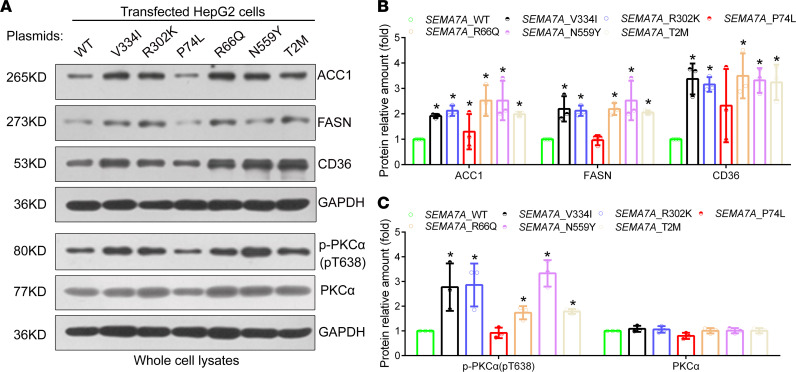
Figure 7. The SEMA7A mutation activates PKC-α signaling in hepatocytes.
HepG2 cells were transfected with the plasmid for the expression of SEMA7A_WT or SEMA7A_V334I, _P302K, _P74L, _R66Q, _N559Y, or _T2M mutant, and the relative levels of ACC1, FASN, CD36, phosphorylated PKC-α (T638), and PKC-α expression in each group of cells were determined by Western blot. (A) Representative images of Western blot analyses. (B) Quantitative analysis of each mutant protein and (C) the relative levels of T638 and PKC-α in HepG2 cells from 3 separate experiments. The levels of each protein in the SEMA7A_WT–transfected cells were designated as 1. The data were analyzed by 1-way ANOVA with Tukey’s post hoc tests or by Kruskal-Wallis test with Dunn’s post hoc test analysis. *P < 0.05 versus the SEMA7A_WT cells.

