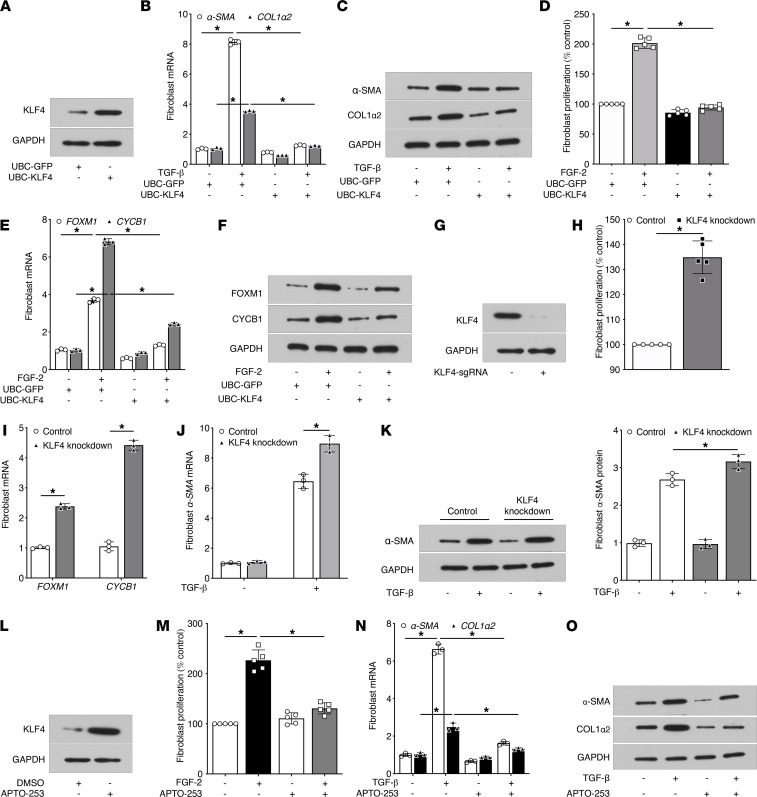
Figure 2. KLF4 expression regulates fibroblast differentiation and proliferation.
(A) KLF4 protein expression in MRC5 cells 48 hours after lentiviral transduction of UBC promoter-driven GFP (UBC-GFP) or human KLF4 (UBC-KLF4). (B and C) Effect of UBC-KLF4 or -GFP on TGF-β–induced expression of α-SMA and COL1α2 mRNA at 24 hours by qPCR (B) and protein at 48 hours by Western blot (C). (D–F) Effect of UBC-KLF4 or -GFP on FGF-2–induced fibroblast proliferation as determined at 72 hours by the CyQuant NF DNA binding assay (D) and proliferation-associated expression of FOXM1 and cyclin B1 (CYCB1) mRNA by qPCR (E) and protein by Western blot (F) at 48 hours. (G) CRISPR-mediated knockdown of KLF4 protein in MRC5 cells as determined by Western blot. (H and I) Effect of KLF4 knockdown on basal proliferation of fibroblasts as determined by the CyQuant NF DNA binding assay (H) and basal expression of FOXM1 and CYCB1 mRNA by qPCR (I). (J and K) Effect of KLF4 knockdown on TGF-β–induced expression of α-SMA analyzed at 48 hours by qPCR (J) and Western blot (left) and its protein densitometry (right) (K). (L) KLF4 protein induction in fibroblasts after treatment with APTO-253 (250 nM) for 36 hours. (M) Effect of APTO-253 on baseline and FGF-2–induced proliferation of fibroblasts as determined by the CyQuant NF DNA binding assay. (N and O) Effect of APTO-253 on TGF-β–induced expression of α-SMA and COL1α2 analyzed at 48 hours by qPCR (N) and Western blot (O). GAPDH mRNA and protein were used to normalize α-SMA, COL1α2, FOXM1, and CYCB1 expression by qPCR and Western blot, respectively. In A, C, F, G, K, L and O, representative Western blot of 2–3 experiments is shown. Data in B, E, I, J and N are expressed relative to control values and are shown as mean ± SEM from 3 independent experiments. *P < 0.05, 2-way ANOVA.

