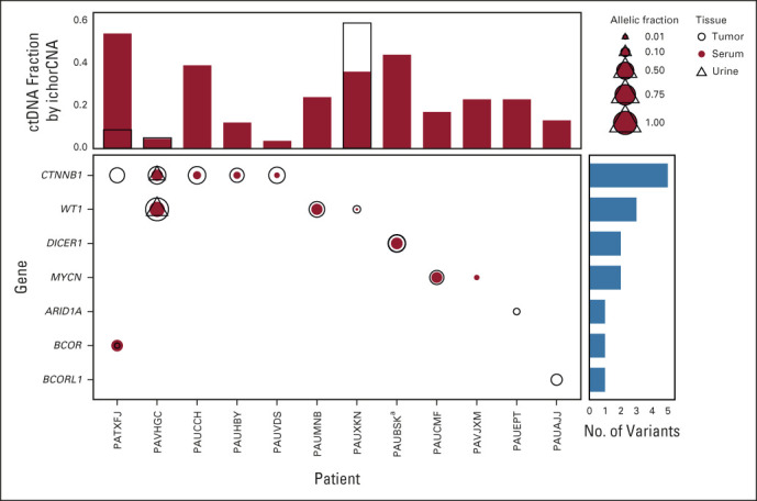
FIG 2.

SNVs identified in tumors and matched ctDNA samples. Plotted are somatic variants identified in genes frequently mutated in WTs. Open circles indicate variants identified in tumors, red circles indicate variants identified in serum, and open triangles identified from urine. The size of the symbols reflects the relative allelic fraction of the identified events. Vertical bar plots reflect the fraction of ctDNA identified in the serum (red bars) and urine (open bars with black outline). Horizontal blue bars summarize the number of variants identified in the indicated gene. aA case with two variants in the DICER1 gene identified in both the tumor and the serum. ctDNA, circulating tumor DNA; SNV, single-nucleotide variant; WT, Wilms tumor.
