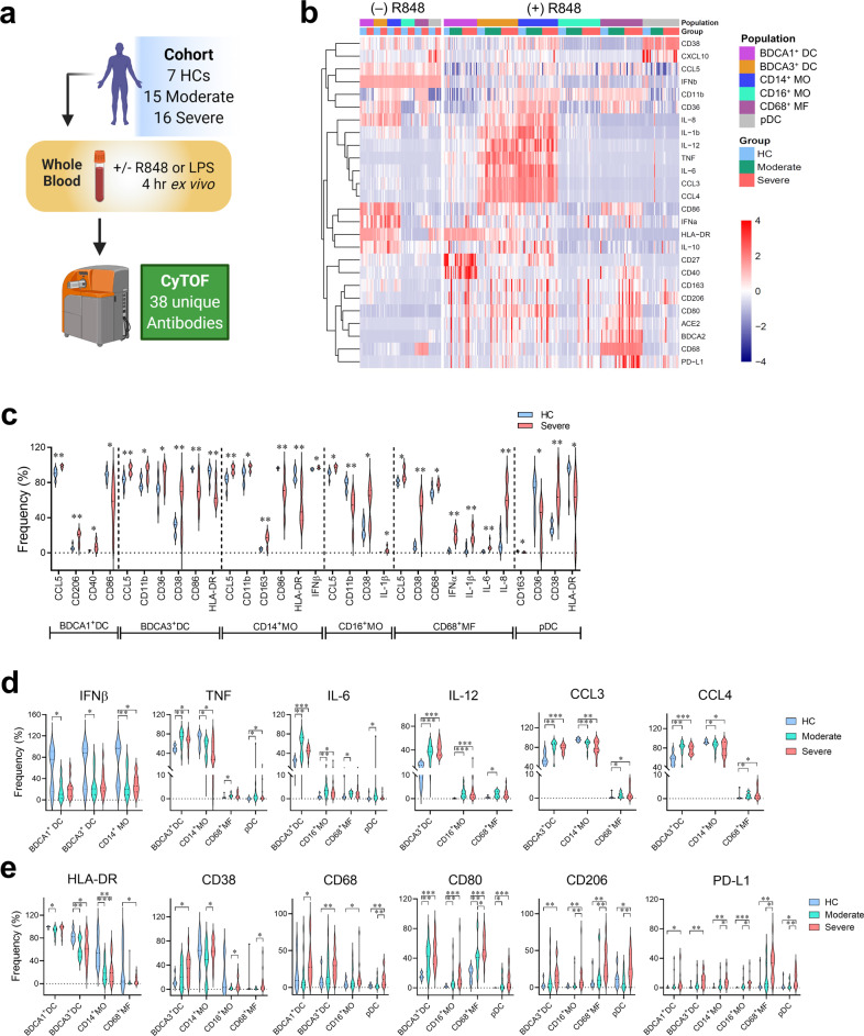
Fig. 5. Hyperinflammatory states of blood myeloid cells from COVID-19 patients before and after ex vivo stimulation with R848.
Whole blood samples from cohort (n = 7 healthy controls (HC); n = 15 moderate COVID-19 patients; n = 16 severe COVID-19 patients) were stimulated ex vivo with or without R848 for 4 h and then stained with 38 metal-conjugated antibodies for mass cytometry (CyTOF) analysis. a Pipeline for processing, treatment, and analysis of blood samples from the cohort. Image generated with BioRender. b Heatmap of the frequencies of peripheral blood myeloid populations expressing indicated markers with and without R848 stimulation. c Violin plots of the frequencies of myeloid populations expressing indicated markers from HC (n = 7) and cells from patients with severe COVID-19 (n = 7) without R848 treatment. d, e Violin plots of frequencies of myeloid populations stimulated ex vivo with R848 expressing indicated cytokines and chemokines (d) as well as surface markers (e) from HCs (n = 7) and COVID-19 patients with moderate (n = 15) or severe (n = 16) diseases. *P < 0.05; **P < 0.01; ***P < 0.001.

