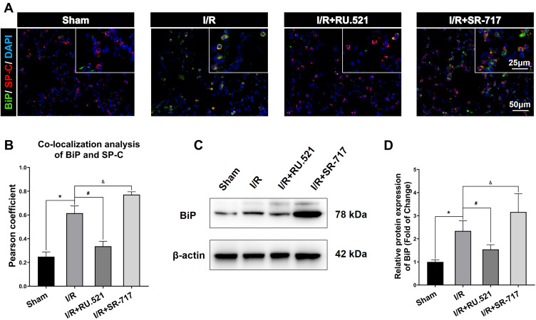
Figure 4.
BiP expression in alveolar epithelial type II cells by using Western blot and double immunofluorescent staining respectively. (A) Double immunofluorescent staining to colocalize BiP with alveolar epithelial type II cells marker SP-C in lung tissue. Scale bar = 25μm or 50μm. (B) The quantified colocalization of BiP with alveolar epithelial type II cells marker was assessed using the Pearson coefficient. Data are presented as mean ± SEM, n = 6 per group. (C-D) Western blotting analysis to verify the expression of BiP. Data are presented as mean ± SEM, n = 3 per group. *P < 0.05 vs sham group, #P < 0.05 vs I/R group, &P < 0.05 vs I/R group.

