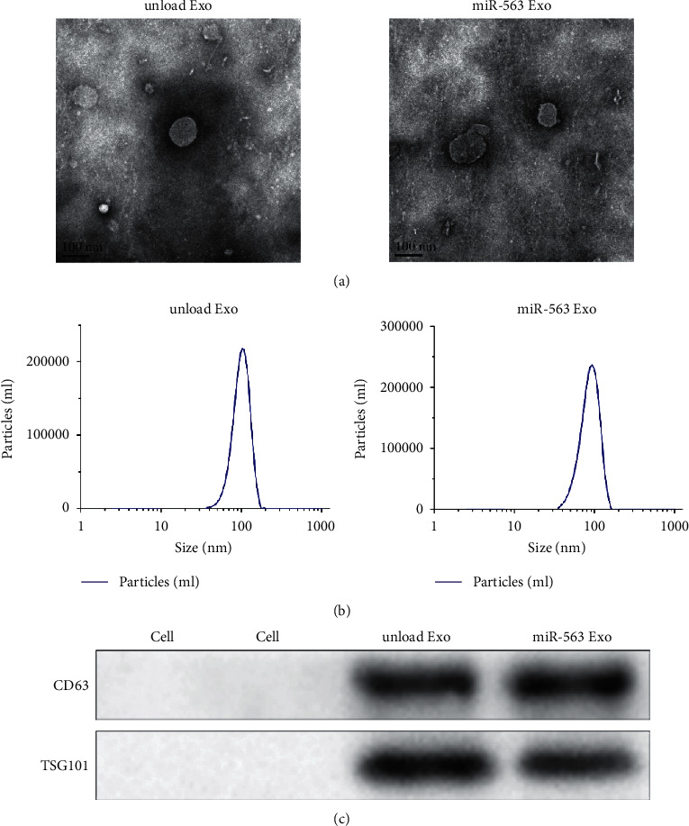
Figure 2.

Characterization of miR-563 loaded exosomes. Transmission electron photomicrographs of the unloaded Exo and prepared miR-563 Exo. (b) Representative image of DLS showing the concentration and size distribution of the unloaded Exo and prepared miR-563 Exo. (c) Western blot analysis of exosomal surface markers CD63 and TSG101.
