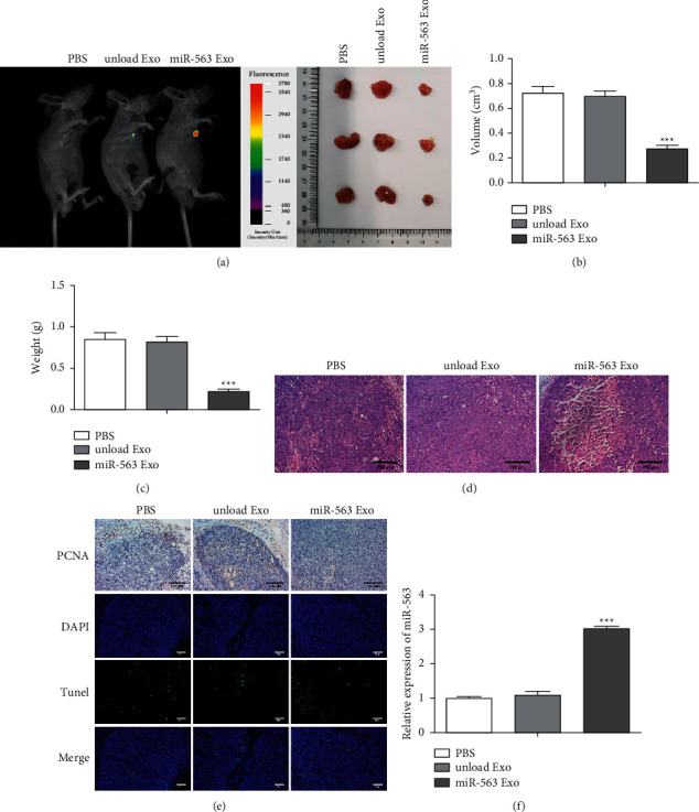
Figure 6.

The antitumor role of miR-563 loaded Exo in vivo. (a) Imaging of PKH26-labeled exosomes in mice. (b) Tumor images in different groups of mice and the changes in tumor volume of mice treated with different treatments with 16 days. (c) Tumor weight of mice in different groups. (d) H&E of tumor sections. Scale bar, 100 μm. (e). TUNEL staining on tumor sections. Scale bar, 100 μm. (f) MiR-563 expression assessed by real-time qPCR. The values were presented as mean ± SD (n = 3). ∗∗∗P < 0.001 versus the unloaded Exo group.
