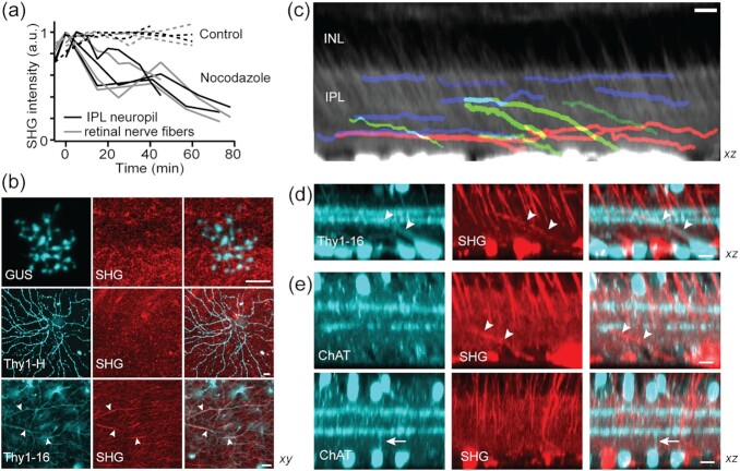
Fig. 6.
The origin of the IPL neuropil signal. (a) SHG intensity after the treatment with nocodazole. (b) Co-registration of SHG and GFP/YFP in the GUS, Thy1-YFP-H, and -16 retinas. Overlaps only in Thy1-YFP-16 (arrowheads). (c) The stratification of mid- (blue), long-range (red), and displaced amacrine cell (green) SHG+ neurites. (d and e) The identity of SHG+ displaced cell. SHG+ neurites are Thy1+ but not ChAT+ (arrowheads). Conversely, the neurites of ChAT cells are SHG- (arrow). Scale bars, 10 µm.

