Fig. 7.
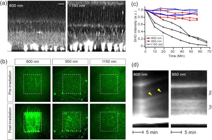
Retinal SHG imaging is tunable to longer wavelengths for safety. (a) SHG imaging at  = 800 and 1150 nm. Scale bars, 20 µm. (b and c) Light-induced changes evaluated by autofluorescence and SHG, respectively. The postirradiation images are the 10th of z-stacks acquired every 5 min (i.e., t = 50 min). (d) SHG kymographs of the retina under continuous illumination. Axial swelling at 800 nm (arrowheads) but not at 950 nm.

