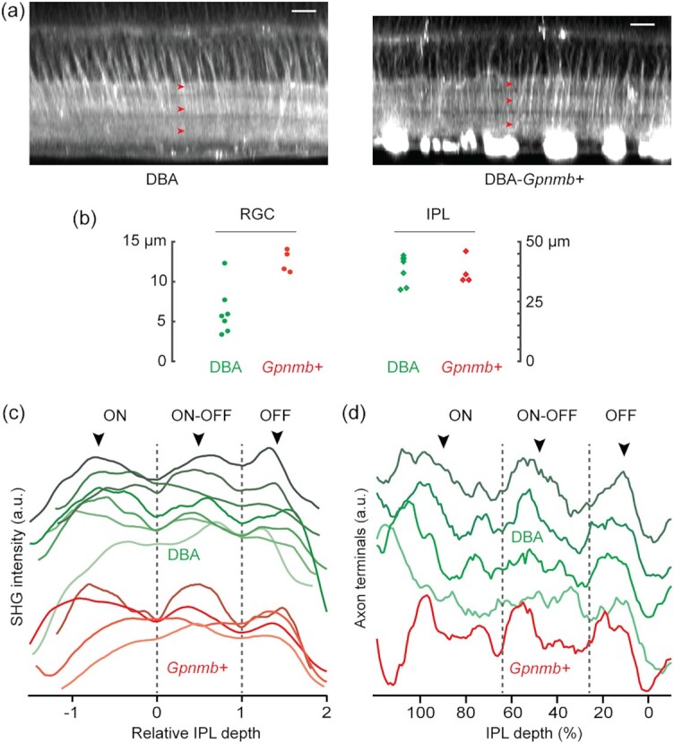
Fig. 8.

Morphological changes in the glaucomatous inner retina. (a) Representative SHG images of DBA and DBA-Gpnmb+. The IPL sublaminae appear normal in both strains (arrowheads). Scale bars, 20 µm. (b) The thicknesses of the RGC axon bundles vs. the IPL. (c) The average SHG intensity of the IPL neuropil. (d) The density of axon terminals of bipolar cells.
