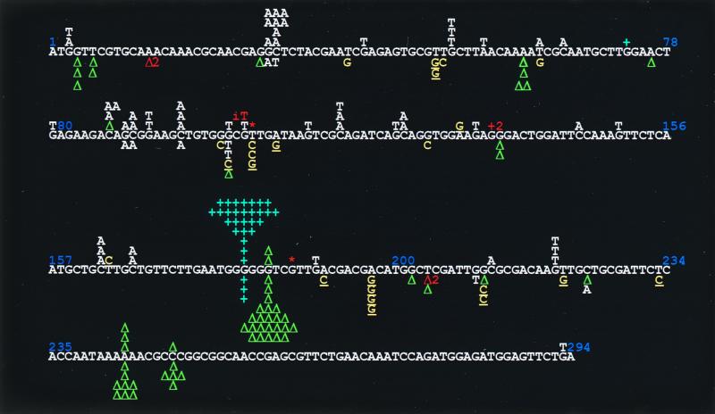
FIG. 1.
cII mutational spectra determined in E. coli AB1157 (wild-type) strain. Mutants recovered in AB1157 transformed with the pWKS30 or pYG782 vector are indicated above or below, respectively, the cII coding sequence. Base substitutions to G:C and A:T are represented in yellow and white, respectively. The G:C-directed substitutions in the sequence of 5′-GX-3′, where X represents the base that is changed to G, is underlined. Blue +, +1 frameshift mutation; green triangle, −1 frameshift mutation. Other mutations are colored in orange: −2 deletion at positions 16 to 18; T insertion (iT) between positions 101 and 102; 84-bp deletion between the GTT direct repeat marked by *; +2 addition at positions 134 and 135 and -CT or -TC deletion at positions 203 to 205. Position 200 is T of a TGG sequence.

