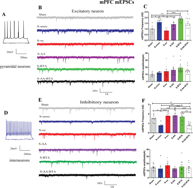
Fig. 7.
SCFA treatment rescued synaptic deficits in layer II/III of the mPFC neurons in cognitively compromised pain rats. A–C Effect of the SCFAs intervention on the spontaneous synaptic transmission of excitatory neurons in pain-induced cognitive deficits rats. A Current-clamp recordings to identify excitatory neurons in the mPFC slices. B Original traces of mEPSCs of excitatory neurons recorded from the six groups. C Bar graphs showing the frequency and the amplitude of mEPSCs in mPFC excitatory neurons. N = 5 rats for each group. D–F Spontaneous synaptic transmission of the mPFC inhibitory neurons in slices. D Current-clamp recordings to identify inhibitory neurons in the mPFC slices. E Original traces of mEPSCs in slices from the six groups. F Bar graphs showing the frequency and the amplitude of mEPSCs in mPFC inhibitory neurons. Results are expressed as mean ± SEM; n = 5 rats for each group. Tukey’s post hoc tests; *p < 0.05, **p < 0.01, ***p < 0.001, ****p < 0.0001

