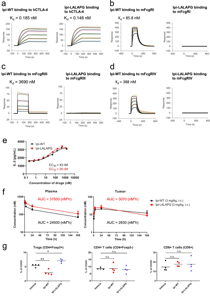
Fig. 3.
Ipilimumab mIgG2a LALAPG exhibits similar binding to human CTLA-4 and similar plasma and tumor PK as ipilimumab mIgG2a wildtype but does not bind to mouse FcγRs or induce in vivo Fc-dependent intratumoral Treg depletion. Surface plasmon resonance measurements of Ipi-WT and Ipi-LALAPG to a human CTLA-4, b mouse FcγRI, c FcγRIII and d FcγRIV. Human CTLA-4 was injected at concentrations ranging from 3.125 to 50 nM, and mouse FcγRs were injected at concentrations ranging from 187.5 to 3000 nM. e Inhibitory activity against interaction between CTLA-4 and B7 ligands by Ipi-WT and Ipi-LALAPG and their EC50 values. PHA-stimulated human primary CD3 + T cells expressing huCTLA-4 were co-cultured with engineered Raji cells expressing CTLA-4 ligands CD80 and CD86 in the presence of Ipi-WT or Ipi-LALAPG. Data are representative of 4 donors. Human-CTLA-4 knock-in C57BL/6 mice were inoculated with 1 × 10⁶ MC38 cells. When the mean tumor volume reached approximately 580 mm3, animals were randomized into treatment groups (n = 3/group) and dosing was initiated. f Plasma (left) and tumor (right) concentration–time profiles and the AUC of Ipi-WT (3 mg/kg) or Ipi-LALAPG (3 mg/kg) in MC38 tumor-bearing huCTLA-4 knock-in C57BL/6 mice (n = 3) via intravenous administration. Mice were treated with Ipi-WT or Ipi-LALAPG at day 0. At indicated time points after single dosing, mice were euthanized and plasma and tumor tissues were harvested. Ipi-WT or Ipi-LALAPG was quantified using ELISA. g Quantification of tumor-infiltrating Tregs, CD4 + T cells and CD8 + T cells. Mice (n = 4) were treated with PBS (vehicle), 3 mg/kg of Ipi-WT or 3 mg/kg of Ipi-LALAPG at day 0. The mice were euthanized 72 h after single dosing, and single-cells were isolated from resected tumors and analyzed by flow cytometry. n.s., nonsignificant. *P < 0.05 and **P < 0.01 by one-way ANOVA with Dunnett’s multiple comparisons test

