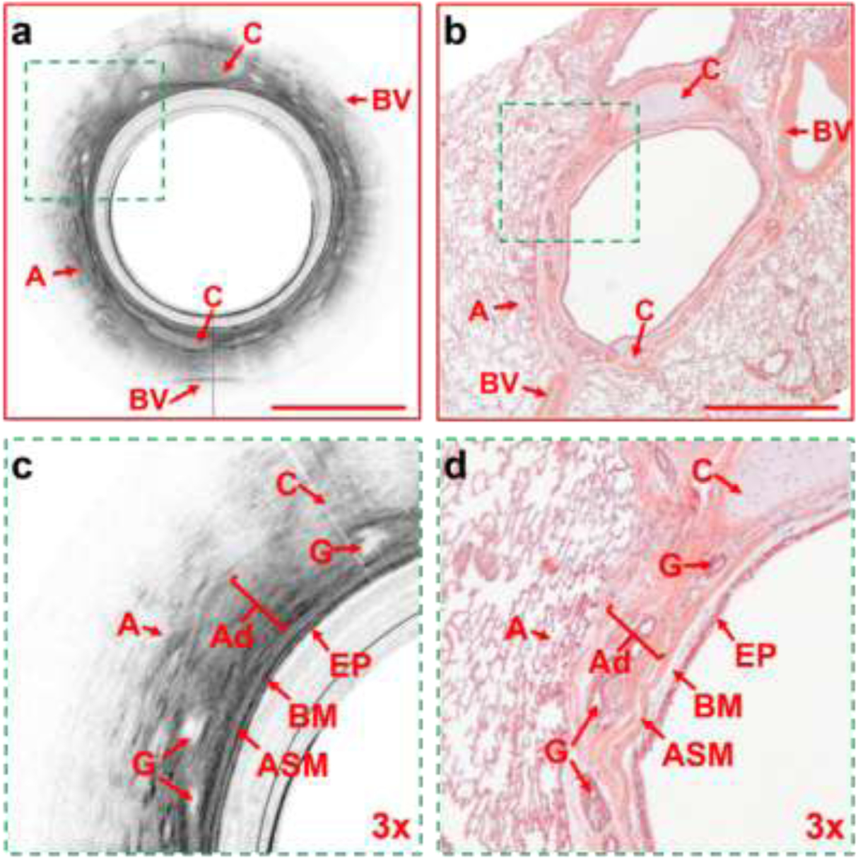
Figure 3:

Representative cross-sectional OCT and histological images. (a) The OCT airway image of the entire matched airway. (b) H & E histological image of the entire matched airway. (c) the 3X enlarged matched area from Figure 3a showing the structural components of the airway wall. (d) the 3X enlarged matched area from Figure 3b showing the matched structural components of the airway wall. A= alveoli, C= cartilage, BV=blood vessel, EP = epithelium, BM = basement membrane, ASM= airway smooth muscle, G = glands, Ad = adventitia. Scale bars: 1 mm.
