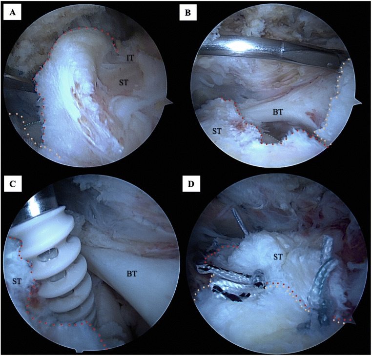
Fig. 7.
Arthroscopic photos of the subacromial space in the lateral decubitus position demonstrating full-thickness supraspinatus tendon (ST) and infraspinatus tendon (IT) tears with a large lateral intact tendon stump (yellow tracing) on the greater tuberosity footprint but an adequate amount of medial tendon (red tracing) at the muscle tendon junction for suture repair, consistent with a type IA tear pattern as classified by Millet et al. [25]. A View of the full-thickness ST and IT tears and the underlying articular cartilage of the humerus (blue tracing) through the anterior portal. B View of the full-thickness ST tear and the biceps tendon (BT) through the posterior portal. C Suture anchors placed in the great tuberosity for ST repair. D Repair of the tendon achieved with 3 suture anchors, demonstrating the large lateral tendon stump (yellow tracing) still intact on the tuberosity

