Fig. 1.
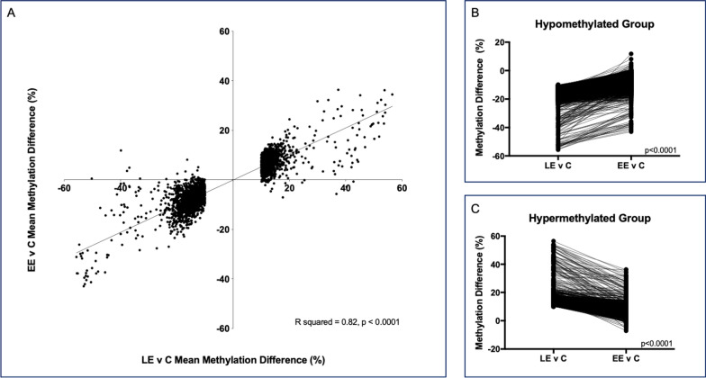
The impact of exposure timing on DNA methylation. A Methylation differences at the top 3321 dmCpG sites between the late exposed (LE) and control animals are plotted on the x-axis. Methylation differences between early exposed (EE) and control animals at those same CpG sites are plotted on the y-axis. Negative values represent hypomethylation relative to controls, while positive values represent hypermethylation relative to controls. Linear regression shows significant agreement between the direction of methylation change across the exposure groups (p < 0.0001, R2 = 0.82). Of the top 3321 dmCpG sites between LE and control animals, we separated those that were hypomethylated relative to controls (B) from those that were hypermethylated relative to controls (C). For B and C, the methylation difference at each CpG site is plotted on the y-axis. The x-axis represents the methylation difference at a specific CpG site between the LE and control animals and the methylation difference at that same CpG site between the EE and control animals, connected by the solid line. This demonstrates that regardless of the direction of methylation change, the magnitude of the mean methylation difference at each CpG site is significantly reduced between EE and control sperm relative to the difference present between the LE and control sperm (p < 0.0001, Bonferroni corrected)

