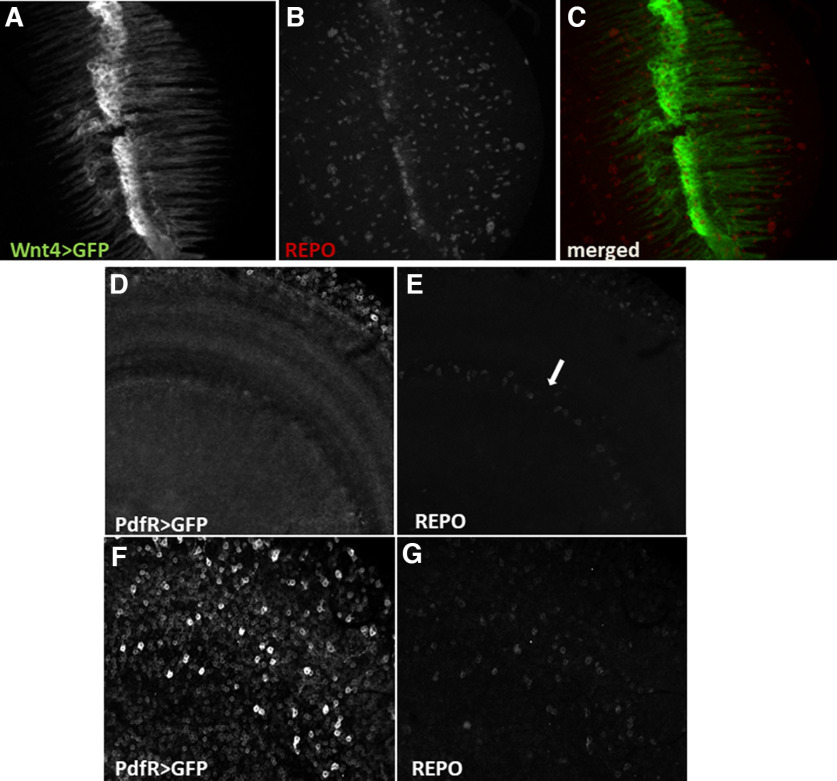
Figure 4.
Chiasm glia oscillators are not regulated by PDF released from LNvs. A–C, Whole-brain immunostaining of Wnt4>GFP flies using anti-Repo antibodies visualized chiasm glia cells (A, C, green) and pan-glial nuclei (B, C, red). D–G, Whole-brain anti-Repo immunostaining of PDFR>GFP flies visualized cells expressing receptor for PDF neuropeptide (D, F) and glial cells nuclei (E, G). D, E, Optical section shows no signal in the place where chiasm glia is located (marked with arrow). F, G, Glial cells other than chiasm glia show expression of PDFR.

