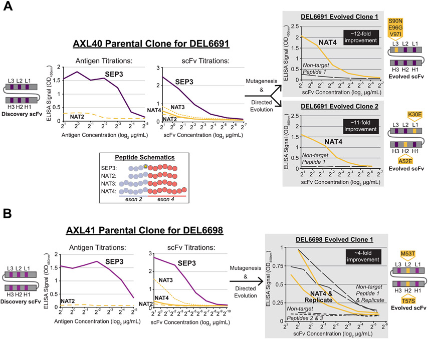
Fig. 2. Successful directed evolution of SEP-specific scFvs to the native exon 2/4 splice junction of CENP-A-ΔExon3.
Unique scFvs from the AXL40 (A, left) and AXL41 (B, left) Discovery screens were purified and tested in antigen and/or scFv titration ELISAs against the SEP and NAT peptides shown in the inset schematic, color coded as per Fig. 1. A total of 10 SEP-specific scFvs were identified (Supplementary Table S2) and two clonal examples are shown here. Note that the scFvs in (A) are unique in both the framework and CDR regions compared to the scFvs in (B), denoted by different shades of grey/purple in the scFv schematics and line graphs. (A and B, right) Unique scFvs from the Directed Evolution screens (DEL6691 and DEL6698) were purified and tested in scFv titration ELISAs against the NAT4 peptide and non-target peptides. Non-target peptides were used as a negative control to assess nonspecific binding events. Three evolved scFvs with 4- to 12-fold improved binding strength to the native exon 2/4 splice junction sequence were identified. Directed Evolution scFv schematics are shown with preserved parental CDRs in purple and mutated CDRs (AA change and position are indicated) in yellow.

