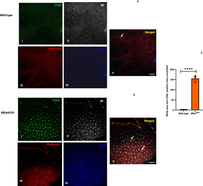
Figure 4.
Confirmation of the microglial origin of autofluorescent spots in Mfrp KI/KI mice retina. (A,B) Retinal wholemount immunostaining for CD68 positive cells and autofluorescent spots using confocal microscopy with × 20 lens (Right panel), enlarged view of individual spots (I-CD68, II-AF (Autofluorescence), III-phalloidin (for actin, staining) IV-DAPI, V-Merged), (Left panel) show that (C) a significantly higher number of autofluorescent spots were present in MfrpKI/KI mice retina (****p < 0.0001) (n = 3).

