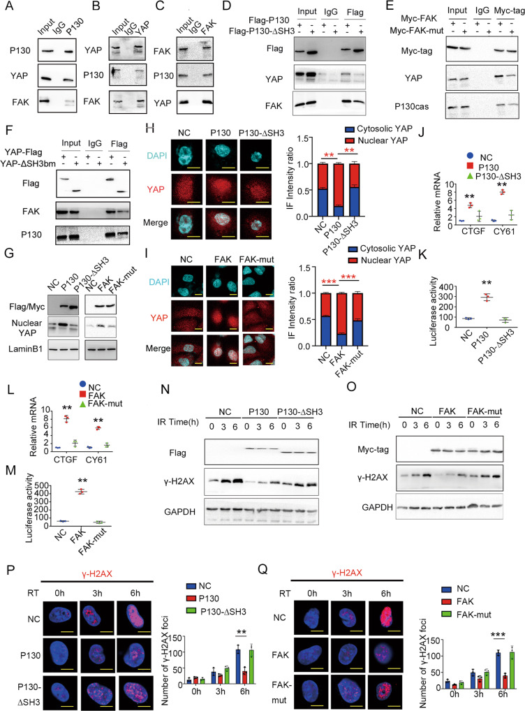
Fig. 5. Interaction with P130cas is pivotal in forming the triple complex with FAK and YAP.
A P130cas was immunoprecipitated from A549 cells and immunoblotted with indicated antibodies. B YAP was immunoprecipitated from A549 cells and immunoblotted with indicated antibodies. C FAK was immunoprecipitated from A549 cells and immunoblotted with indicated antibodies. D P130cas was immunoprecipitated by Flag antibody from A549 cells infected with Flag-P130cas lentivirus vector or Flag-P130-ΔSH3 lentivirus vector and immunoblotted with indicated antibodies. E FAK was immunoprecipitated by Myc antibody from A549 cells transfected with Myc-FAK or Myc-FAK-mut (712/715A) and immunoblotted with indicated antibodies. F YAP was immunoprecipitated by Flag antibody from A549 cells transfected with Flag-YAP or Flag-YAP-ΔSH3bm and immunoblotted with indicated antibodies. In A549 cells overexpressing Flag-P130cas/Flag-P130-ΔSH3 or overexpressing Myc-FAK/Myc-FAK-mut (712/715A), after nucleoplasmic separation, using immunoblotting to evaluate P130cas/FAK, nuclear YAP and LaminB1 (G), using immunofluorescence (H, I) to identify the subcellular distribution of YAP, using qPCR assay (J, L) and luciferase reporter assay (K, M) to detect the downstream gene activity of YAP (t test. *P < 0.05). In A549 cells overexpressing Flag-P130cas/Flag-P130-ΔSH3 or overexpressing Myc-FAK/Myc-FAK-mut (712/715A), using immunoblotting to evaluateγ-H2AX and GAPDH at the indicated time points after 5 Gy ionizing radiation (N, O), representative immunofluorescence images of the number of γ-H2AX foci at the indicated time points after 5 Gy ionizing radiation (P, Q, scale bar = 10 μm). Each experiment was quantified as Mean ± SD of three independent experiments (t-test, two-sided, **P < 0.01, ***P < 0.001). For Western blot experiments, the samples derive from the same experiment and the gels/blots were processed in parallel.

