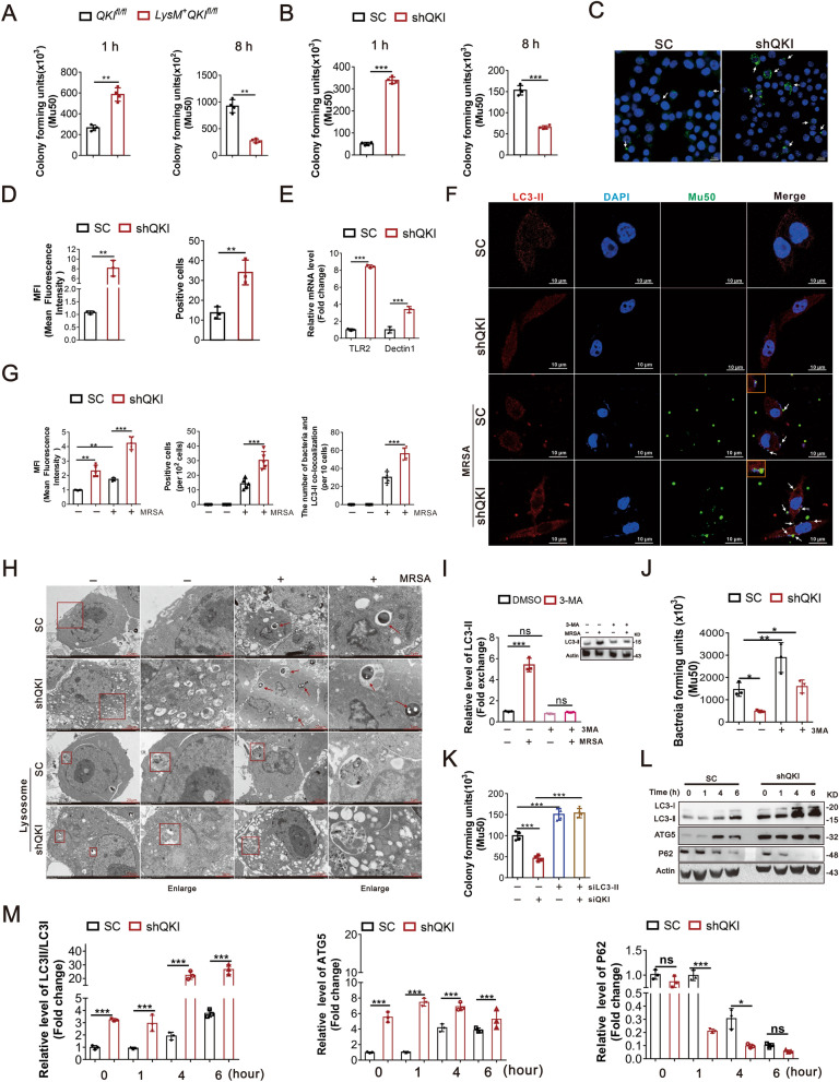
Fig. 2.
QKI deficiency improved the phagocytic and bactericidal activity of macrophages in vitro. A After peritoneal cells were harvested from QKIfl/fl and LysM + QKIfl/fl mice, F4/80+CD11b+ macrophages were isolated by FACS and incubated with MRSA. At 1 h or 8 h post-incubation, the number of bacteria units forming was counted respectively. Enumeration of forming bacteria units (c.f.u) were shown in the left (1 h) and right (8 h). B RAW264.7 cells transfection with shRNA specific for QKI and scrambled shRNA control were incubated with MRSA. The number of bacteria units forming in RAW264.7 cells were calculated and shown after indicated time. C Phagocytosis ability was analyzed by phagocytose opsonized particles in RAW264.7 cells after incubated with Latex beads-rabbits IgG-FITC complex. Representative graph of positive area was showed. D Graphs showed the quantification of mean fluorescence intensity (MFI) and number of FITC staining positive cells in three randomly fields. E Real-time PCR analysis of relative mRNA expression of the receptors level (TLR2 and Dectin-1) in QKI silenced (shQKI) and scramble (SC) RAW264.7 cells after incubating with MRSA. F Confocal microscopic imaging of co-localization of FITC-conA stained Mu50 and LC3-II in QKI silenced (ShQKI) and scramble (SC) RAW264.7 cells after MRSA infection. FITC-conA stained Mu50: green, immuno-stained for LC3-II: red, DAPI: blue. (Scale Bar: 10 μm). G The quantification of mean fluorescence intensity (MFI) of LC3-II immunostaining was showed (left). The number of FITC staining positive cells in five randomly fields (middle). The number of bacteria and LC3-II co-localization in 10 cells. Five fields were randomly selected to analyzed. H The formation of autophagosomes in QKI silenced (ShQKI) and scramble (SC) RAW264.7 cells were observed under transmission electron microscopy. Bacteria within autophagosomes were indicated with thick arrow (Scale Bar: 2 μm). The lysosomes were observed in SC and ShQKI cells, which showed in square box. I Western blots were performed to determine the effects of autophagy inhibitor 3-MA with or without MRSA infection. One representative immunoblot is shown (on the left) and the graph (on the right) presents quantification of the band intensity for immunoblots from three independent experiments. The results were shown as expression of LC3-II. J “Phagocytosis assay” was used to determine the effect of 3-MA to phagocytosis ability of QKI silenced (ShQKI) and scramble (SC) RAW264.7 cells after treated with MRSA and 3-MA simultaneously. The number of bacteria units forming were calculated after 8 h. Enumeration of forming bacteria units (c.f.u) were shown. K LC3-II was knockdown by siRNA, follwed by “Phagocytosis assay” determining the effect of LC3 to phagocytosis ability of QKI silenced (ShQKI) and scramble (SC) RAW264.7 cells. L Immunoblot analysis of protein expression of LC3, ATG5 and P62, in scramble and shQKI RAW264.7 cells, with actin as an internal control. One representative immunoblot is shown. M Graphs are representative quantification of the band intensity for immunoblots for figure L, which was from three independent experiments. All the bars represented the mean of measurements from three independent experiments, and the error bars indicated ± SD. (A, B, J and L) are representative of one experiment (A and B, n = 4 wells/group; J and L, n = 3 wells/group). (D) n = 3 fields/group. (E) are representative of one experiment (n = 3 wells/group). (D) n = 5 fields/group. (b and L) are representative of three experiments. *p < 0.05, **p < 0.01. ***p < 0.001, not significant (ns) (student’s t test)

