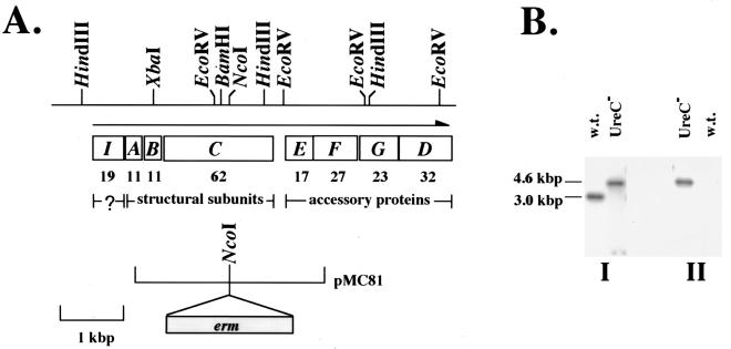
FIG. 1.
Construction and characterization of UreC-deficient S. salivarius. (A) A restriction map of the chromosomal region containing the ure cluster is shown at the top. The organization of the operon is shown, and the direction of transcription is indicated by a horizontal arrow. The molecular mass (in kilodaltons) of each open reading frame is shown below the gene. The limits of pMC81 in relation to the ure cluster and the location of erm within pMC81 are also shown. (B) Southern blot analysis of the wild-type strain and otherwise isogenic ureC-deficient derivative. Total cellular DNA was digested with HindIII and transferred onto two membranes simultaneously by a sandwich blot. The membranes were probed with a ureC-specific probe (I) or with an erm-specific probe (II).

